Programs & Services
CAF Invests in Technology to Help Military Personnel with an OSI
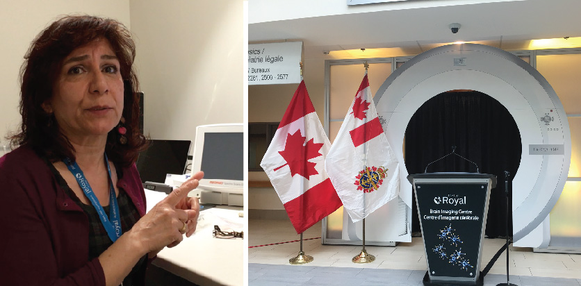
Last month, the Canadian Armed Forces (CAF) announced a momentous partnership with the Royal Ottawa Mental Health Centre that entails the use of brain imaging technology. This brain imaging technology in itself is momentous and combines two different medical imaging techniques for the first time.
The Positron Emission Tomography (PET scan)/ Functional magnetic resonance imaging (fMRI) is the first of its kind in Canada with the sole purpose of researching mental health. The Royal has stated that through this machine doctors will be able to better study mental health in order to provide personalized treatment to soldiers diagnosed with Post Traumatic Stress Disorder (PTSD) and other Operational Stress Injuries (OSIs).
“With this scanner we have, we can measure all those sorts of chemicals in the brain. There are other scanners, and there have been studies about the different chemicals of any different part of the brain but [with] this scanner especially we have extra hardware/software for multi-nuclear detection. So, we can basically detect more chemical properties than any other scanner,” said Dr. Fakhereh Mirrashed, manager of the PET/fMRI.
Mirrashed explained that the scanner combines the functions of an MRI with nuclear medicine imaging to pinpoint exact locations in the brain for a better prognosis.
“Of course, we have MRI, very advanced field in medical imaging. We have nuclear medicine, very advanced in medical imaging, but these were not married together until we had the technology to put these together into one casket and they are not interfering with each other to distort,” explained Mirrashed.
Previously, researchers and doctors had to use MRIs and nuclear imaging separately. Since each image individually did not always show a complete picture, doctors had to superimpose the images. This forced the medical community to make an educated guess as to exact locations in the body with irregularities. For example, when studying mental health researchers are not looking for a tumour or something that can be physically taken out, making it difficult for an MRI alone to pick up.
“What we are looking for with mental health is at the cellular, molecular level. What is this neurotransmitter doing, what sort of the chemicals are they back and forth communicating, and of course, those give us the behaviour we have, the action that we do, the emotion we feel. Those are all that we are going to measure right here on this scanner,” added Mirrashed.
Research can now be taken to another level with the combination of the nuclear medicine and the MRI. Patients can be injected with an isotope tracer that will travel through the blood stream and lodge in the part of the brain where cells and molecules are functioning irregularly.
“…[The researcher] sends [the isotope] as a GPS, basically to go through your body and get the cellular that aren’t visible in the MRI because they aren’t showing any big sized tumour but they’re doing something they aren’t supposed to do,” stated Mirrashed.
The scanner can then print an exact image that includes the nuclear medicine technology with the MRI image. After this, images can be sent for data processing and results can be handed to doctors to decide the best course of treatment based on the chemicals in the brain, as shown by the scanner images. The scanner can be used for follow-up to see if the treatment is working.
Mirrashed explains with this technology patients will be spared the try and see approach when it comes to medication. With an image from the machine, doctors will be able to see what medications are working for a patient and which ones are not.
Watch the full video here:


